Human Tissue Laboratory
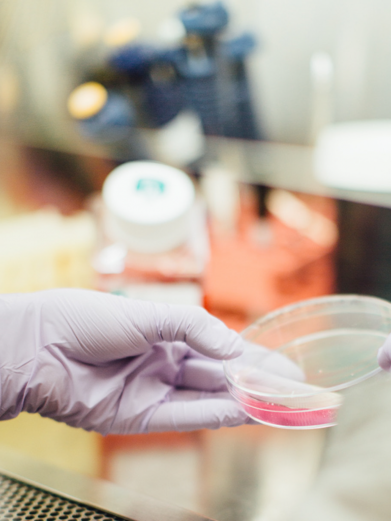
To support the development of novel therapies in Neuroscience and Neuro-oncology we need access to human tissue such as adult and fetal human brain tissue, blood and Cerebrospinal Fluid (CSF).
Supplementary to our Human Tissue Lab, we also work across two sites in Cardiff and Swansea to support Neurological Biobanking and tissue phenotyping. Learn more about Biobanking
What is HTL used for?
- Scale up human cell and tissue models for studying disease
- Mechanisms
- Generate novel therapies
- Validate targets and signalling changes
We have set up a unique human adult tissue facility to perform 2D and 3D culturing of primary brain tissue for disease modelling
Where is the tissue collected from?
The tissue used for research is obtained by collecting excess tissue removed from brain surgery. Surgeries include:
- Epilepsy Surgery
- Tumour Surgery
- Head injury surgery
What is the tissue used for?
We use cultures of human brain to study how the brain works and what goes wrong in disease. We look at:
- Primary research on neurological disease in Epilepsy, traumatic brain injury (TBI) and oncology
Translation and validation of mechanisms and potential therapies from animal studies in:
- Immune modulation
- Cell therapy
- Oncolytic Viral Therapies
- Gene Therapy Delivery
- Lipid Biosynthetic Pathway Analysis
Learn more about
BioBanking at BRAIN

Case studies
-
CASE STUDY 1: Culturing Human Brain tissue in 3D
We are using a unique method for culturing human brain tissue to study disease mechanisms and test new therapies. These cultures are 3-dimensional and capture the heterogeneity of human brain cells. This allows us to research different neurological diseases (e.g. epilepsy, traumatic brain injury and cancer) and model the individual response to potential therapies.

Figure 2: Low power 3D view of cultures showing nerve cells, glia and immune cells

Figure 3: GFAP+ Astrocytes (green) allow us to study glial cells

Figure 4: Immune cells – microglia in cultures Helps us look at the inflammation in the brain
-
CASE STUDY 2: Early Diagnoses of Brain Infection
Using Cerebrospinal Fluid (CSF) samples from patients with suspected infection we are examining the use of immune markers to accurately diagnose infection much earlier than with standard microbiology cultures and so treat patients earlier.
Collaboration with Prof M Eberl, Dr S Cuff, Dr J Merola & Prof W Gray.

Figure 4: Immune cells – microglia in cultures Helps us look at the inflammation in the brain
-
CASE STUDY 3: Providing Embryonic Stem (ES) and Fetal Tissue to model and validate cell replacement therapies
Our human fetal (hF) tissue collections are unique in the UK in providing high quality hF brain tissues for research and clinical application. We have used this tissue for critical preclinical cell replacement therapy (CRT) research, understanding neurodegenerative and developmental mechanisms and underpinning CRT clinical trials.
Our work provides:
- HTA Standard hF donor cells for clinical CRT trials (RfPPB funded TRIDENT)
- Research grade hF tissue to improve cell survival, circuit repair and functionality, and optimize delivery methodology.
- Research grade hF cells as gold-standard validation for developing hES and hiPS derived CRTS
- Research grade hF cells for disease modelling both in vitro and in vivo.

Figure 7: Studying the electrical excitability of human nerve cells. Human stem cell-derived neuron progenitors have been considered as a potential candidate for cell replacement therapy. The green-labelled human nerve cells started to gain electrical activity after being co-cultured with human astrocytes for 2 weeks. Increased firing rate was observed in those neurons 4 weeks after the co-culture. This model will be further optimised to mimic the pathological microenvironment and investigate its impact on transplanted neuron progenitors for human studies.
