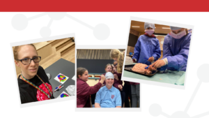
May 2023 was a busy month for the BRAIN Unit, from science talks science to hosting the highly anticipated BRAIN games, with primary school attendees from across Cardiff. Read on to find out more.
Science with a pint: hearts and minds research
On May 5th, BRAIN researchers Professor Anne Rosser and Dr Emma Lane hosted a ‘Hearts and Minds’ event in Radyr Golf Club. Attendees had the opportunity to hear about the latest research and clinical developments in neurological diseases and cardiology and take part in demonstrations. The night saw some amazing talks, including one from Dr John Huish who talked about the history of cardiology. Interventional radiologist, Chris Williams brought a range of stents for attendees to try and deploy, as well as rat brain sections which were stained to reveal dopaminergic transplants – displayed under a microscope!
Exploring the effects of samba on neuroplasticity in Parkinson’s disease
May 13th saw Parkinson’s researchers from Bangor, Cardiff and Swansea come together to host simultaneous events, along with the Wales Research Interest group. One of the events was funded by the Dementia Research Institute and the BRAIN involve group, both based in the Hadyn Ellis Building (HEB) at Cardiff University. Around 100 people with Parkinson’s and their family members came to HEB to hear about the latest research in Parkinson’s disease and see the launch of an all-new samba group for people with Parkinson’s: ‘SParky Samba’.
‘SParky Samba’ aims to develop strong community bonds between those with lived experience of Parkinson’s as well as explore the benefits of Samba percussion in promoting neuroplasticity. It is facilitated by a local Cardiff-based Afro Brazilian percussion band named Barracwda. You can find out more about SParky Samba by following them on Twitter.
Promoting equality and diversity in research
Dr Emma Lane was invited to speak at two events hosted by the Equality Diversion Inclusion in Research (EDIRA) project group on 12th and 19th May. EDIRA aims to create an inclusive research framework, developing guidance using the perspectives of underserved communities and supporting the work of professionals from various Communities of Practice.
Another year, another BRAIN Games for primary pupils
On May 19th, 4 Cardiff primary schools visited two of Cardiff University’s research facilities: Spark and CUBRIC to learn more about brain research and take part in a variety of fun brain-related activities.
Researchers at Cardiff University have been awarded funding that will enable them to better map the brain to treat diseases such as epilepsy, dementia, and multiple sclerosis.
A £1 million grant from the Medical Research Council was secured by Cardiff University, along with University College London, Leeds and the University of Cape Western Reserve. The Cardiff University team includes Professor Derek Jones, Professor Liam Gray, Khalid Hamandi and Dr Marco Palombo.
The grant will enable researchers to ‘make the invisible visible’ by obtaining high-quality images of the human brain and learning the mapping between Magnetic Resonance Imaging (MRI) and histology, the microscopic study of human tissue.
Making the invisible visible
Professor Liam Gray explained, “One of the key challenges in diagnosing brain disease such as epilepsy, dementia or multiple sclerosis is the difficulty in detecting small and subtle cortical abnormalities that are not easily identified. Conventional MRI scanners can detect abnormal signals, but it’s impossible to tell what’s driving them; this could be differences in cell size, shape or density. Currently, such information can only be obtained by cutting the tissue and examining it under a microscope.”
Examining the cortex using state-of-the-art equipment
The funding will enable researchers to take advantage of advances in MRI physics, which hold the promise of detecting and characterising tissue abnormalities that are currently “invisible”. Such technologies have been applied to the examination of white matter, which is associated with psychiatric diseases such as schizophrenia, but the cortex remains unexplored to an extent.
To illuminate infected tissue, BRAIN researchers will scan patients in state-of-the-art equipment, remove pathological tissue through surgery, transport it to an experimental MRI scanner for extended scanning, and then light microscopy and electron microscopy will be used. These techniques allow us to see the brain over a variety of scales of magnification.
This will allow us to establish an MRI signalling ‘fingerprint’ of specific disease processes in the cortex that are currently invisible to conventional MRI, creating a significant change in our ability to localise pathologies in the brain and monitor them non-invasively over time.
Funding research opportunities
The grant will also fund an 18-month Clinical Research Fellow in an Experimental Functional Neurosurgery position. The Clinical Research Fellow will be responsible for preparing and recruiting study participants as well as dealing with and processing surgical tissue during the study.
Professor Gray will carry out surgery to extract tissue samples from patients, who will be anonymously linked to the work so data can remain confidential. It is then processed in the Human Tissue Laboratories of the BRAIN Unit at the University Hospital of Wales (YAC). Samples will then be sent to colleagues on the MRC grant who will scan the tissue and process it for Immunohistochemistry (IHC) and electron microscopy which will show disease evidence that will be mapped to the Magnetic Resonance Imaging (MRI).
Professor Liam Gray said, “If successful, this technology could help us localise the cortical pathology that causes epilepsy in the brain and allow surgery to be more accurately targeted to cure epilepsy. It could also significantly expand the population of patients suitable for surgery.”
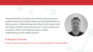
Dr Benjamin Dummer is a Research Assistant based in a lab at the University Hospital of Wales (UHW), under the supervision of Professor Liam Gray.
The research
Our current research focuses on glioblastoma, which is a deadly brain cancer. We are characterising a 3D cell culture model using brain tumour tissue direct from surgery. This will allow us to personalise medicines to individual patients and gain a better understanding of glioblastoma biology.
We are also collaborating with other research groups within Cardiff University and outside institutions to explore how our 3D model can be used to better understand other neurological diseases.
Despite decades of research and millions of pounds spent, patient outcomes for people diagnosed with glioblastomas is still very poor. There is a clear need for developing novel treatments to combat the disease. Within these tumours there is a subpopulation of cells called stem cells which are implicated in treatment resistant and disease progression. Understanding how these cells interact with each other and their microenvironment is a fascinating area of research which will hopefully lead to a better understanding of this deadly disease.
What next?
Our next step is to complete the work on our 3D model so we can move forward to developing a personalised medicine paradigm with the aim of helping patients who suffer with this horrible disease.